Categories
Microbiology

Study reveals role of hormone in heart disease risk

Scientists at the University of Veterinary Medicine, Vienna have shed light on the role of phosphate-rich foods in raising blood pressure and promoting vascular calcifications.

Levels of a hormone known as Fibroblast Growth Factor 23 (FGF23) are increased as a result of a high-phosphate diet and this puts a strain on the cardiovascular system. 

Foods that are high in phosphates include processed cheese, Parmesan, cola, baking powder and most processed foods. Phosphates are also frequently used in the food industry as preservatives and pH stabilizers.

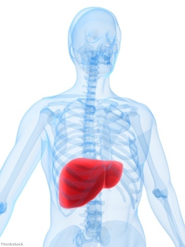
Chronic kidney disease affects more than 500 million people around the world. Clinical studies have shown that such people are prone to developing cardiovascular diseases such as high blood pressure and vascular calcification.

However, until recently, the link between renal disease, the build-up of the hormone FGF23, which is produced in the bones, and cardiovascular disease was unclear.  

The scientists demonstrated that FGF23 controls the reabsorption of filtered sodium in the kidneys. Mice lacking this hormone excrete higher quantities of sodium in their urine, resulting in low blood pressure. 

Those with high levels of FGF23, however, show increased levels of sodium in their blood, and in turn, high blood pressure.

Increased levels of FGF23 place a strain on the heart. "In patients with chronic renal disease, both the phosphate levels and the levels of FGF23 are chronically high. This often leads to cardiovascular disease," said Reinhold Erben, the head of the Unit of Physiology, Pathophysiology and Biophysics at the Vetmeduni Vienna.

A second study reveals that FGF23 controls calcium levels in the blood. Too much of the hormone encourages the kidneys to take up calcium, leading to vascular calcification.

FGF23 is formed in the bones and controls the excretion of phosphate via the kidneys. When abnormally high levels of phosphate are found in the body, FGF23 levels rise, leading to the excretion of excess phosphate.

If the excretion process via the kidneys does not work correctly, or too much phosphate is taken in with food, phosphate and FGF23 levels increase, resulting in a spiral which could have damaging health consequences.

Categories
Microbiology

UA scientists make liver disease breakthrough

Scientists at the University of Arizona (UA) have made a breakthrough in the study of end-stage liver disease that could help to develop new treatments for the condition.

End-stage liver disease goes hand-in-hand with oxidative stress – damage to body tissues caused by reactive oxygen molecules or free radicals, which occur naturally as a result of the body's metabolic processes. 

A number of mechanisms are employed by cells to keep these free radicals under control, most of which involve a protein known as Nrf2. This activates biochemical processes that capture reactive oxygen molecules or dispose of damaged cellular components before they can cause more trouble.

When no oxidative stress response is needed, an enzyme known as Keap1 keeps levels of Nrf2 low.

According to conventional wisdom, conditions of oxidative stress lead the body to activate the  the Nrf2-mediated protection pathway in order to limit the damage caused by the destructive oxygen compounds. However, contrary to expectations, this mechanism is not activated during liver cirrhosis. 

Donna Zhang, a professor in the UA Department of Pharmacology and Toxicology, said "This was a puzzle before we did our study. Somehow the protective mechanism mediated by Nrf2 is compromised by another factor, other than Keap1, in liver cirrhosis."

Additionally, drugs aimed at inhibiting Keap1 from chewing up Nrf2 have proven ineffective in a cirrhotic liver.

A study of tissue samples revealed the reason behind the observation. Another enzyme, known as Hrd1, breaks down Nrf2 and prevents the much-needed antioxidant response, exacerbating the disease process.

Hrd1 – part of cells' waste disposal mechanism –  specialises in destroying misfolded proteins before they can accumulate and damage cell components.

Levels of Hrd1 are low under normal conditions so it does not interfere with Nrf2. But as liver cirrhosis progresses, excessive inflammation triggers the waste-mediated stress response, Hrd1 becomes very abundant and begins chewing up Nrf2.

New therapeutic treatments could be developed as a result of the scientists' insights. The team were able to restore Nrf2 levels in cirrhotic liver tissue by inactivating Hrd1 in experiments, effectively reversing liver cirrhosis in mice.

Categories
Microbiology

Gene therapy developed to reverse memory loss in mice

Researchers from the Institute of Neuroscience at the Universitat Autonoma de Barcelona (UAB) have developed a gene therapy that could pave the way for new Alzheimer's disease treatments.

One reason why attempts to devise a treatment have hitherto been unsuccessful is a lack of knowledge on the cellular mechanisms which cause alterations in nerve transmissions and the loss of memory in the initial stages of the disease.

However, the UAB researchers have discovered the cellular mechanism behind memory consolidation. Their gene therapy was able to reverse memory loss in mice models with the initial stages of the disease.

They achieved this by injecting into the hippocampus a gene which causes the production of a protein blocked in patients with Alzheimer's, the "Crtc1" (CREB regulated transcription coactivator-1).

The protein thus restored gives way to the signals needed to activate the genes involved in long-term memory consolidation.

In order to identify the gene, gene expression in the hippocampus of healthy control mice was compared with that of transgenic mice which had developed the disease.

The team used DNA microchips to identify the genes and proteins which expressed themselves at different stages of the disease. 

It was found that those genes involved in memory consolidation coincided with the genes regulating Crtc1, a protein which also controls genes related to the metabolism of glucose and to cancer. Altering this group of genes could cause memory loss in the initial stages of Alzheimer's.

The formation of amyloid plaque aggregates, which cause Alzheimer's, prevents the proper functioning of the Crtc1 protein.

"When the Crtc1 protein is altered, the genes responsible for the synapsis or connections between neurons in the hippocampus cannot be activated and the individual cannot perform memory tasks correctly," explained Carlos Saura, researcher of the UAB Institute of Neuroscience and head of the research. 

Mr Saura said new perspectives on therapeutic prevention have been opened up by the research, as they have demonstrated the effectiveness of gene therapy in preventing the loss of memory in mice.

Alzheimer's disease affects almost 500,000 people in the UK, making it the most common form of dementia.

Categories
Microbiology

Scientists discover role of protein in combating cancer

Researchers have identified a new 'dustbin' role for a molecule that helps a drug to kill cancer cells.

The new study, published in the journal Proceedings of the National Academy of Sciences on Monday April 21st, could be used to develop a test to identify patients who would be most responsive to a new class of cancer drugs and those who may develop resistance, as well as suggesting new approaches to discovering more effective drugs.

Researchers found that a molecule, known as Cullin-5 (CUL5), acts as a combined cleaner and dustman, sweeping up proteins that tell cancers to divide continuously and consigning them to a cellular 'dustbin'.

CUL5 works in opposition to another important molecule known as HSP90, which scientists have been trying to block with drugs to prevent cancer cells from dividing.

When the scientists treated cancer cells with drugs that block HSP90, they found that the cleaning protein CUL5 stepped in to dispose of the proteins that were telling the cells to keep dividing.

They also discovered an additional function of CUL5, showing that it helps to pull the 'dividing-signal' proteins away from the protective shelter of HSP90, effectively stopping cancer in its tracks by labelling the cells with a tag that sends them to the cellular dustbin.

Professor Paul Workman, study lead and deputy chief executive of The Institute of Cancer Research, said drugs that block HSP90 have previously been identified as a potential treatment for cancers and the team knew CUL5 may play a role in these drugs' function.

"Our new research shows that CUL5 is not only vital in the response of cancer cells to HSP90 inhibitors but also reveals surprising insights into precisely how it works by acting at several different levels," he added.

Professor Workman said he was surprised by the high number of cancer-causing proteins CUL5 helps to dispose of and the fact that it works across several types of tumour.

The researchers said patients may be resistant to HSP90-blocking drugs if their cells have low levels of CUL5, while the drugs may work better in those with high levels of the molecule. 

Categories
Microbiology

DNA looping damage ‘could play role in HPV-related cancers’

Researchers at the Ohio State University have shed light on the way in which certain strains of human papillomavirus (HPV) cause cancer.

They found that the virus disrupts the human DNA sequence with repeating loops when it is inserted into host-cell DNA as it replicates.

Around 610,000 cases of cancer per year are caused by HPV. This accounts for about five per cent of all cases of the disease and virtually all incidences of cervical cancer. The mechanisms behind the process are not yet completely understood, however.

The team of scientists utilised the immense computational power of the Ohio Supercomputer Center to carry out their research. 

A variety of techniques, including whole-genome sequencing and genomic alignment, were used to examine ten cancer-cell lines and two head and neck tumour samples from patients.

David Symer, assistant professor of molecular virology, immunology and medical genetics at Ohio State's Comprehensive Cancer Center, said: "HPV can act like a tornado hitting the genome, disrupting and rearranging nearby host-cell genes. 

"This can lead to overexpression of cancer-causing genes in some cases, or it can disrupt protective tumour-suppressor genes in others. Both kinds of damage likely promote the development of cancer."

Co-senior author Professor Maura Gillison explained that they observed fragments of host cell genome being removed, rearranged or increased in number at sites of HPV insertion.

These were accompanied by increases in the number of HPV copies in the host cell, while the expression of cancer-promoting genes viral E6 and E7 was also increased.

Although E6 and E7 are essential for the development of cancer, they are not alone sufficient to cause it. Researchers believe the destabilising loops may play a key role, providing additional alterations that promote cancer development, as genomic instability is a hallmark of cancers.

Professor Symer said the team's research shows what happens during the 'end game' of cancer development. He added that it sheds light on the steps between initial infection with a HPV to the development of a related cancer.

Categories
Microbiology

Study identifies possible bowel cancer treatment target

New research reveals the role played by microRNA in the development of colorectal cancer and shows it could be a key target for treatments.

A study into the role played by the molecule in bowel cancer was carried out by an international team which included scientists based at The Institute of Cancer Research, London, the University of Glasgow and Ohio State University in the US.

Their findings are published in the journal Cancer Cell, with funding for the UK research team coming from the Kimmel Cancer Foundation, Cancer Research UK, The Marie Curie Actions Programme and a Scottish Senior Clinical Research Fellowship.

Scientists discovered that MicroRNA 135b is employed by a number of cancer genes to drive the development of the disease.

They believe drugs targeted at the molecule could eliminate the effects of multiple cancer-causing mutations, while tests for it could identify those with the most aggressive form of the disorder.

An analysis of 485 people with bowel cancer found that levels of microRNA 135b were at least four times as high in tumours as in healthy tissue. Patients with the highest amount of the microRNA survived for the least amount of time.

A study conducted on mouse models found that blocking the micro-RNA stopped tumour growth. In half of the animals, tumours regressed so dramatically that they could no longer be seen by imaging, while the animals displayed no side-effects.

Additionally, a number of known cancer gene variations, such as APC, PI3KCA, SRC and p53, were found to exercise their effects through microRNA 135b.

This is significant because many people develop a resistance to treatments blocking bowel cancer mutations. Inhibiting microRNA 135b could attack cancer without resistance occurring by blocking the effects of multiple cancer-causing mutations simultaneously.

Testing for microRNA 135b could also identify patients who are most in need of treatment.

Professor Owen Sansom, deputy director of The Cancer Research UK Beatson Institute at Glasgow University, said: "This exciting work shows a single microRNA can control multiple pathways that go wrong in colon cancer and offers the hope that in the future this could be targeted in patients that have colon cancer."

Categories
Microbiology

Micro-RNA provides insight into hereditary trauma

Researchers have gained a new insight into how traumatic experiences can affect successive generations of animals.

It has long been known in psychology that traumatic experience can induce behavioural disorders that can be passed down from one generation to the next but the underlying physiological mechanisms are poorly understood.

Isabelle Mansuy, professor at ETH Zurich and the University of Zurich, has been studying the molecular processes involved in non-genetic inheritance of behavioural symptoms induced by traumatic experiences in early life.

Her team has identified micro-RNA molecules as key factors in these processes. Enzymes synthesise these RNAs from specific sections of DNA and they are then trimmed by other enzymes into mature forms.

Cells naturally contain a number of micro-RNAs, which have a number of regulatory functions, such as controlling how many copies of a particular protein are made.

The researchers studied the number and kind of micro-RNAs expressed by adult mice exposed to traumatic conditions in early life and compared them with non-traumatised mice.

It was discovered that stress changes the amount of several micro-RNAs in the blood, brain and sperm. 

A comparison with the tissues or cells of control animals found that some micro-RNAs were produced to excess, while the others were under-produced. This resulted in a misregulation of cellular processes normally controlled by these molecules.

The mice behaved markedly differently following their traumatic experiences, exhibiting depressive-like behaviour and partly losing their natural aversion to open spaces and bright light.

While the offspring of the mice were not directly affected by traumatic stress, these behavioural symptoms were passed on to the next generation via sperm.

The metabolism of these stressed mice was also affected: their insulin and blood sugar levels were found to be lower than in the offspring of non-traumatized parents, and these even persisted three generations down the line.

It is an open question how the misregulation in short RNAs occurs, but Professor Mansuy said it probably begins with the overproduction of stress hormones.

She said other traits may also be passed on through similar mechanisms. The research team is currently investigating the role of short RNAs in trauma inheritance in humans.

Categories
Microbiology

Bone marrow stem cells ‘could help to treat stroke victims’

A research team from UC Irvine's Sue & Bill Gross Stem Cell Research Center has found that stem cells derived from bone marrow may have the potential to treat stroke victims.

Neurologist Dr Steven Cramer and biomedical engineer Weian Zhao conducted an analysis of published research, identifying 46 studies that used mesenchymal stromal cells (MSCs) in animal models of stroke.

MSCs, which are a type of multipotent adult stem cells mostly processed from bone marrow, were found to be significantly better than control therapy in 44 of the studies.

Significantly, the effects of the cells on recovery were strong irrespective of the dosage, the time the MSCs were administered relative to stroke onset or the method of administration.

MSCs were effective whether introduced via the blood or the brain, even if they were administered a month after the event.

"Stroke remains a major cause of disability, and we are encouraged that the preclinical evidence shows [MSCs'] efficacy with ischemic stroke," said Dr Cramer, a professor of neurology and leading stroke expert. 

"MSCs are of particular interest because they come from bone marrow, which is readily available, and are relatively easy to culture. In addition, they already have demonstrated value when used to treat other human diseases."

While MSCs transform into a wide variety of cell types, such as bone, cartilage and fat cells, they do not differentiate into neural cells.

The cells nevertheless play important roles in promoting brain repair following a stroke. They are attracted to injury sites and release a wide range of molecules in response to signals released by these damaged areas.

MSCs are responsible for a number of activities: blood vessel creation to enhance circulation, protection of cells starting to die and growth of brain cells, among others.

When MSCs are able to reach the bloodstream, they gather in parts of the body that control the immune system and help to create an environment that is more conducive to brain repair.

Dr Cramer said the findings could form the basis of further studies on the use of MSCs in the treatment of ischemic stroke in humans.

Categories
Microbiology

New immune system discovery ‘could help treat cancer’

A discovery that adds a new drug target to cancer immunotherapy has been identified by researchers from La Jolla Institute for Allergy and Immunology and their collaborators from other institutes.

The study reveals a new way to block the function of immune inhibitory checkpoint receptor CTLA-4, which is already generating a large amount of interest in the pharmaceutical and research communities due to its cancer-fighting potential.

Led by Dr Amnon Altman and Dr Kok-Fai Kong, the team demonstrated a previously unknown interaction between CTLA-4 and an intracellular enzyme protein kinase known as C-eta.

This interaction is critical for the functioning of regulatory T cells, which suppress the immune system. 

While this activity is normally a useful part of a healthy immune system, preventing the body from potentially damaging immune responses that lead to autoimmune diseases, it can inhibit beneficial immune attacks against cancer.

CTLA-4 is a protein that is found on the surface of regulatory T-cells, where it plays an important role in immune suppression.

"The way it works is that this enzyme physically binds to the CTLA-4 receptor," said Dr Altman. "This binding is critical for certain suppressive functions of the regulatory T cells to proceed."

Dr Altman and his team found the enzyme must bind to CTLA-4 in order for the regulatory T cells to turn down the immune system in mice.

They also discovered that eliminating this enzyme prevented regulatory T cells from suppressing the immune system's response against a growing tumour.

Although regulatory T cells lacking protein kinase C-eta failed to inhibit an immune response against a growing tumour, they were able to continue to inhibit autoimmune disease in a mouse model of inflammatory bowel disease.

"This means that you could potentially create a therapy that would allow for a more effective immune response against cancer without the risk of increasing susceptibility to autoimmune diseases," said Dr Altman. 

He added that this is important as it points to an immune mechanism specific to tumours which does not result in a response that could lead to autoimmune diseases.

Dr Altman said this is possibly due to the fact that regulatory T cells use diverse means to suppress different immune responses that do not depend on the link between C-eta and CTLA-4.

Categories
Microbiology

Researchers grow self-healing muscle

Biomedical engineers have successfully grown living skeletal muscle that has many properties of ordinary muscle tissue and, for the first time, demonstrates its ability to heal itself in the laboratory and in animals.

Researchers at Duke University tested the bioengineered muscle by watching it through a window on the back of a living mouse. This allowed them to monitor the muscle's integration and maturation inside a living, walking animal.

Nenad Bursac, associate professor of biomedical engineering at Duke, said: "The muscle we have made represents an important advance for the field. It's the first time engineered muscle has been created that contracts as strongly as native neonatal skeletal muscle."

Throughout years of honing their research methods, a team led by Prof Bursac and graduate student Mark Juhas found two things are necessary for preparing better muscle: well-developed contractile muscle fibres and a pool of muscle stem cells, known as satellite cells.

Satellite cells are available to every muscle, ready to activate upon injury and begin the regeneration process. The key to the team's success lay in creating the microenvironments – called niches – where these stem cells await their 'call to duty'.

The researchers subjected the muscle to a number of laboratory trials. They measured its contractile strength by stimulating it with electrical pulses and showed it was more than ten times stronger than any previously engineered muscles. 

A toxin found in snake venom was used to prove the satellite cells could activate, multiply and successfully heal the injured muscle fibers.

The muscle was then inserted into a small chamber on the backs of mice which was covered by a glass panel. They checked on its progress every two days for two weeks.

Muscle fibres had been genetically modified to produce fluorescent flashes during calcium spikes – which cause muscle to contract – and researchers were able to watch the flashes grow brighter as it strengthened. 

"We could see and measure in real time how blood vessels grew into the implanted muscle fibres, maturing toward equalling the strength of its native counterpart," Mr Juhas commented.

The team are now beginning to undertake work to see whether their bioengineered muscle can be used to repair actual injuries and treat disease.