Categories
Microbiology

Chromosomal translocations reproduced in human cells

For the first time, scientists have reproduced tumour chromosomal translocations within cells associated with two types of cancer – a development that could lead to new therapeutic interventions.

Researchers at the Spanish National Cancer Research Centre (CNIO) and the Spanish National Cardiovascular Research Centre (CNIC) made the breakthrough. The chromosomal modifications they produced are genetically identical to those observed in leukaemia and other types of human cancer.

Modifications that lead to tumour development occur due to multiple changes in cell physiology and specifically in the cell genome. 

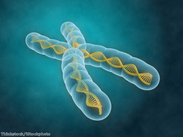
Exchanges of large DNA fragments occur between different chromosomes in leukaemia and other sarcomas. These are known as chromosomal translocations and are necessary for the generation and the progression of a number of neoplastic processes.

The team used RNA-Guided Endonuclease (RGEN) technology or CRISPR/Cas9 genome engineering technology in order to produce these translocations, reproducing them in human stem cells derived from blood and mesenchymal tissue that are identical to those observed in patients with acute myeloid leukaemia or Ewing's sarcoma.

"With this breakthrough it is possible to generate cell models with the same alterations as observed in tumour cells from patients, which will allow us to study their role in tumour development," says CNIO researcher Sandra Rodriguez-Perales. 

"In this way, it will be possible to experimentally recapitulate the necessary subsequent steps for normal cells to transform into cancer cells."

The RGEN tool was developed in 2013 for gene manipulation in eukaryotic cells, including human ones.

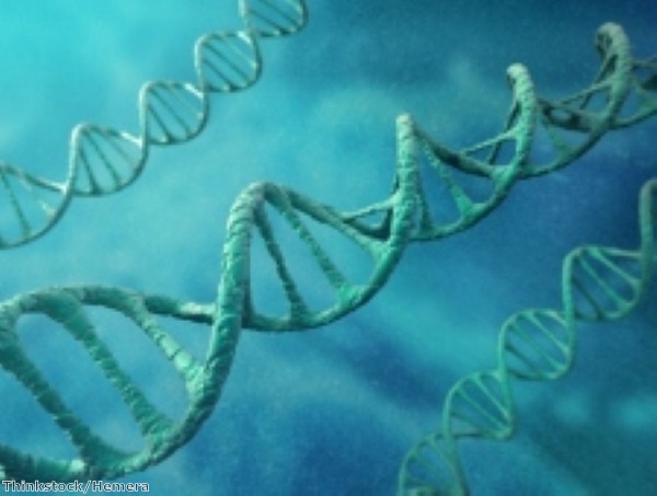
It is based on the design of a small RNA (RNAsg) that is complementary and specific to a 20 nucleotide DNA region.

RNAsg binds to the DNA double helix and acts as a signal for the Cas9 enzyme to make a cut on the edge of the marked DNA. This allows researchers to make cuts on the DNA double helix wherever it is necessary to do so.

Transferring the RGEN components into primary human cells enabled regions of the exchanged chromosomes in some tumours to be marked, thus generating cuts in those chromosomes.

The team claims that better understanding the process of chromosomal translocation will lead to the development of therapeutic interventions.

Categories
Microbiology

Researchers uncover structure of neuronal receptor

Researchers have obtained an unprecedented view of a type of brain cell receptor that is implicated in a range of neurological disorders, including  Alzheimer's disease, Parkinson's disease, depression, schizophrenia, autism, and ischemic injuries associated with stroke.

The team of scientists from Cold Spring Harbor Laboratory say the atomic-level picture of  the intact NMDA (N-methyl, D-aspartate) could prove useful in the development of therapeutic compounds. Their work is published in the journal Science.

NMDA receptors play an essential role in communication between different neurons, integrating both chemical and electrical signals. Such communication forms the basis of memory, learning, and thought, and critically mediates brain development. Increased and decreased NDMA activity is associated with a range of neurological disorders.

A technique known as X-ray crystallography was used to determine the structure of the intact receptor. Numerous interactions between the sub-units of the receptor were identified and the researchers gained new insights into how the complex is regulated.

Protein purification methods were used to isolate the intact receptor, the structure of which was found to resemble a hot air balloon. The 'basket' of the balloon is the transmembrane domain, which forms an ion channel that allows electrical signals to propagate through the neuron

Ion channels act as a gate in a neuronal membrane. When they are closed, ions – electrically charged atoms – are unable to pass through and gather outside cells. When they open, ions travel in and out of cells, generating an electric current that travels through the neuron.

Currents cannot jump between neurons and rely on neurotransmitters, which are triggered by the electrical pulse, to travel the distance between neurons and bind to receptors, such as the NMDA receptor, on the surface of neighboring cells.

Neurotransmitters unlock the ion channels within the receptor and propel the electrical signal across another neuron and, ultimately, across the brain.

The 'balloon' section of NDMA is found outside the cell and binds to neurotransmitters. The structure of the multi-subunit receptor complex helps to explain some of the existing data about how NMDA receptors function.

The team were able to see how one domain on the exterior side of the receptor – the amino terminal domain – directly regulates the ion channel within the membrane and why this is specifically important for the activity of the NMDA receptor.

Categories
Microbiology

New evidence of genetic cause of schizophrenia

Scientists at the Columbia University Medical Center have shed new light on the genetic factors behind an individual's risk of developing schizophrenia.

According to the research, which is published in the latest edition of Neuron, the overall number and nature of mutations – rather than the presence of one single mutation – influence an individual's risk of developing the disease, as well as its severity.

The team sequenced the "exome"- the region of the human genome that codes for proteins – of 231 schizophrenia patients and their unaffected parents. They were able to use the data to demonstrate that collective damage across several genes gives rise to schizophrenia.

Previous research into the disorder has sought to identify individual genes that might act as triggers, but the availability of high throughput DNA sequencing technology has contributed towards a more holistic approach to schizophrenia.

Sequencing data was used to look for genetic differences and identify new loss-of-function mutations in cases of schizophrenia that had not been inherited from the patients' parents. Such mutations are less common, but they have a more severe effect on ordinary gene function.

An excess of loss-of-function mutations was found in a variety of genes across different chromosomes. 

The team also studied what types of mutations are commonly passed on to schizophrenia patients from their parents and found that many of these are "loss of function" types. These were found to occur more frequently in genes with a low tolerance for genetic variation. 

Deeper examinations of the sequencing data were conducted in order to determine the biological functions of the disrupted genes involved in schizophrenia. Two damaging mutations were observed in a gene known as SETD1A, suggesting it contributes significantly to the disease.

SETD1A is involved in a process called chromatin modification – a crucial cellular activity that packages DNA into a smaller volume so it can fit into cells and that regulates how genes are expressed.

The result fits with a body of evidence that damage to chromatin regulatory genes is a common feature of various psychiatric and neurodevelopmental disorders.

Categories
Microbiology

Research sheds light on DNA editing process

An international team of scientists has made significant progress in understanding how enzymes 'edit' genes, which could enable genetic diseases to be corrected.

Researchers at the Universities of Bristol, Munster and the Lithuanian Institute of Biotechnology have studied the process through which enzymes known as CRISPR bind and alter the structure of DNA.

These enzymes were first identified in the 1980s as an immune defence used by bacteria against invading viruses. More recently, it has been shown that one type of CRISPR – Cas9 – can be used to edit the human genome.

CRISPR enzymes have been tailored to target a single pair of letters within the three billion base pairs that make up the DNA molecule. This has been likened to correcting one misspelt word in an encyclopedia of 23 volumes.

In order to accomplish this, the enzymes use a molecule of RNA, which is similar in structure to DNA. The enzymes are required to pull the DNA strands apart and insert the RNA to form a sequence-specific structure known as an R-loop.

Modified microscopes were used to test the R-loop model. DNA models were stretched in a magnetic field and by changing the altering force on the DNA, researchers were able to directly monitor R-loop formation events by individual CRISPR enzymes.

As a result of this technique, the scientists were able to uncover previously hidden steps in the process and to investigate the influence of the sequence of DNA bases.

Professor Mark Szczelkun, from Bristol University's School of Biochemistry, said it is particularly challenging to ensure only one specific area in a genome is targeted when using the genome-editing tools.

"Our single molecule assays have led to a greater understanding of the influence of DNA sequence on R-loop formation," he added.

"In the future this will help in the rational re-engineering of CRISPR enzymes to increase their accuracy and minimise off-target effects. This will be vital if we are to ultimately apply these tools to correct genetic diseases in patients."

Categories
Microbiology

Scientists identify potential diabetes treatment

Scientists have discovered a molecule that slows the degradation of insulin in animals and has the potential to be used as a new treatment for diabetes.

The compound works by inhibiting insulin-degrading enzyme (IDE). Inhibiting IDE in mice elevates insulin levels and promotes insulin signaling in vivo. Researchers at Harvard University say this could be used to maintain higher insulin levels, improve glucose tolerance and therefore to treat diabetes.

Until now, scientists have been unable to regulate the degeneration of insulin and have relied on other methods to treat the disease, such as injecting insulin into diabetic patients.

DNA-templated synthesis, a method for creating new molecules that self-assemble according to an attached DNA sequence, was used to identify the molecule that inhibits IDE.

This system works by combining DNA "templates" – short segments of DNA – with the chemical building blocks of molecules, each of which has its own piece of DNA. When the segments are brought together, the building blocks combine and react, forming more complex molecules.

Sequencing the their DNA strands enables the identification of the resulting molecules.

Researchers incubated the DNA-linked compounds with IDE, hoping that some might bind to the enzyme.

Professor of Chemistry and Chemical Biology David Liu said they hypothesised IDE's activity could be modulated by molecules that were retained by IDE.

"In this case, right out of the library, we found quite a potent and selective inhibitor. Perhaps most important, this molecular had a good half-life in animals, so it could be used to answer the 60-year-old question of what happens when you slow down the natural degradation of insulin in the body," he added.

Experiments with mice showed the compound was able to regulate blood sugar levels. It was found that IDE degrades two other important glucose-regulating peptide hormones – glucagon and amylin – as well as insulin.

Professor Liu said it may be some time before the compound becomes available in pharmacies, as a number of tests and developments need to be made. However, he added that the experimental tools have been put in place for new therapies to be created.

Categories
Microbiology

Handful of building blocks ‘could create many medicines’

Researchers at the University of Illinois (U of I) have identified a new method of synthesising a wide range of medicines using a small number of building blocks.

Thousands of compounds in a class known as polyenes – many of which could be used to develop new drugs – can be constructed simply and economically from a dozen different building blocks.

"We want to understand how these molecules work, and synthesis is a very powerful engine to drive experiments that enable understanding," said Martin Burke, a chemistry professor at the U of I and the Howard Hughes Medical Institute. "We think this is a really powerful road map for getting there."

One simple reaction is used to bind the chemical building blocks, in the same way in which plastic building blocks fit together because they have a simple connector. Scientists thus have greater freedom to construct molecules that may be difficult to extract from their natural source or to synthesise in a laboratory.

Researchers are able to omit or substitute parts of substances in order to create products that may have therapeutic potential. 

Recently, Professor Burke's group synthesised a derivative of the anti-fungal medication amphotericin and gained an insight into how this clinically vital but highly toxic medicine works. They were also able to create a derivative which is an effective fungicide but is nontoxic to human cells.

The team moved on to focus on polyenes and discovered that many elements are common across numerous compounds. They calculated that more than three-quarters of all natural polyene frameworks could be made with only 12 different blocks.

In order to demonstrate their findings, they synthesised a number of compounds representing a wide variety of polyene molecules using only the dozen designated building blocks. 

It is hoped that the identification of the building blocks and their widespread availability will lead to a greater understanding of polyene natural products and their potential as pharmaceuticals, particularly compounds that have hitherto proved to be too costly or time-consuming to make.

Categories
Microbiology

HIV research breakthrough could lead to new therapies

Researchers have uncovered direct proof of a long-suspected cause of multiple HIV-related health complications, supporting the use of complementary therapies to antiretroviral drugs to significantly slow HIV progression.

Scientists at the University of Pittsburgh Center for Vaccine Research (CVR) found that a drug often administered to patients undergoing kidney dialysis significantly reduces the levels of bacteria that escape the gut and reduces health complications in non-human primates infected with the simian form of HIV.

Inflammation and chronic activation of the immune system are major determinants of progression of HIV infection to AIDS. They also play a key role in inducing excessive blood clotting and heart disease in HIV patients.

It was theorised that microbial translocation, which occurs when bacteria in the gut escapes into the body through intestinal lining damaged by HIV, lay behind these findings – but no direct proof of the mechanism existed.

The University of Pittsburgh researchers gave the drug Sevelamer, which is used to treat elevated levels of phosphate in the blood of patients with chronic kidney disease, to monkeys infected with simian immunodeficiency virus, or SIV, the primate form of HIV.

Gut bacteria bind to Sevelamer and this makes it more difficult for them to escape into the body and cause serious problems, such as heart disease, while further weakening the immune system and allowing HIV to progress to full-blown AIDS.

Levels of a protein which indicates microbial translocation remained low in SIV-infected monkeys treated with Sevelamer, while it increased nearly four-fold a week after infection in untreated monkeys.

In treated monkeys, lower levels of a biomarker associated with excessive blood clotting were recorded, demonstrating that heart attacks and stroke in HIV patients are more likely associated with chronic immune system activation and inflammation, rather than HIV drugs.

"These findings clearly demonstrate that stopping bacteria from leaving the gut reduces the rates of many HIV comorbidities," said Dr Ivona Pandrea, professor of pathology at Pitt's CVR.

"Our study points to the importance of early and sustained drug treatment in people infected with HIV."

Categories
Microbiology

Stem cell research sheds light on schizophrenia origins

New stem cell research suggests indicators of schizophrenia are present in early neuron differences, supporting the theory that the risk of developing the disease may begin in the womb.

Scientists at the Salk Institute have uncovered evidence of strange behaviour in the early development stages of neurons generated from the skin cells of people suffering from schizophrenia.

The findings of the study, published in Molecular Psychiatry, suggest the origins of the disorder may lie in the brains of babies still in the womb.

Studying schizophrenia has until recently proved very difficult for scientists, as they have had to examine the brains of patients after death. However, the brains of these patients had often been damaged by age, stress, medication or drug use.

Stem cell technology enabled the Salk scientists to overcome these hurdles. They took skin cells from patients, encouraged them to revert back to an earlier form and then prompted them to grow into very early-stage neurons (dubbed neural progenitor cells or NPCs), which are similar to the cells in the brain of a developing fetus.

The researchers carried out tests on NPCs from the skin cells of four patients with schizophrenia and six people without the disease. 

In one test, they looked at how far the cells moved and interacted with particular surfaces; in the other, they looked at stress in the cells by imaging mitochondria.

The results of both tests demonstrated that NPCs from people with schizophrenia differed in significant ways from those taken from unaffected people.

Unusual activity was detected in two major classes of proteins in cells predisposed to schizophrenia: those involved in adhesion and connectivity and those involved in oxidative stress.

Cells derived from patients with schizophrenia tended to have aberrant migration (which may result in the poor connectivity seen later in the brain) and increased levels of oxidative stress (which can lead to cell death).

These results appear to support the theory that events occurring during pregnancy can contribute to schizophrenia, even though the disorder does not tend to manifest until early adulthood.

"The study hints that there may be opportunities to create diagnostic tests for schizophrenia at an early stage," said Fred Gage, Salk professor of genetics.

Categories
Microbiology

Researchers make personalised medicine breakthrough

Researchers have made a breakthrough in the field of personalised medicine by growing functioning human heart tissue carrying an inherited cardiovascular disease.

The advance comes as the result of a collaborative effort between scientists from the Harvard Stem Cell Institute, the Wyss Institute for Biologically Inspired Engineering, Boston Children's Hospital, the Harvard School of Engineering and Applied Sciences, and Harvard Medical School.

Scientists modelled the cardiovascular disease Barth syndrome, a rare X-linked cardiac disorder caused by mutation of a single gene called Tafazzin, or TAZ.

Skin cells were taken from two Barth patients; these were manipulated to become stem cells that carried the patients' TAZ mutations.

Rather than being used to generate single heart cells in a dish, the cells were tricked into joining together in the way they would if they were forming a diseased human heart. This was achieved by growing them on chips lined with human extracellular matrix proteins that mimic their natural environment.

The tissue thus created contracted very weakly, as would the heart muscle seen in Barth syndrome patients.

Genome editing was then used to mutate TAZ in normal cells, confirming that this mutation is sufficient to cause weak contraction in the engineered tissue.

"You don't really understand the meaning of a single cell's genetic mutation until you build a huge chunk of organ and see how it functions or doesn't function," said Dr Kevin Parker.

"In the case of the cells grown out of patients with Barth syndrome, we saw much weaker contractions and irregular tissue assembly."

The scientists also found that the TAZ mutation works in such a way to disrupt the normal activity of mitochondria, which produce energy – although the overall energy supply of the cells appeared to be unaffected by the mutation.

They describe a direct link between mitochondrial function and a heart cell's ability to build itself in a way that allows it to contract – a newly identified function for mitochondria.

Barth syndrome cells produce an excess amount of reactive oxygen species or ROS as a result of the TAZ mutation. In the laboratory, quenching ROS production restores contractile function, but the scientists have yet to ascertain whether this could be replicated in human or animal models.

Categories
Microbiology

Study casts doubt on heart stem cell regeneration

A new study challenges the efficacy of using stem cells to regenerate damaged heart tissue.

Researchers at Cincinnati Childrens Hospital Medical Center and the Howard Hughes Medical Institute (HHMI) claim that cardiac stem cells used in ongoing clinical trials – which express a protein marker called c-kit – do not achieve high enough regeneration rates to justify their use.

Many patients have already been treated using c-kit positive stem cells, which are removed from healthy regions in a damaged heart, processed in a laboratory and then re-injected into the patients' hearts.

This is an experimental treatment based on preclinical studies in rats and mice which have indicated that c-kit-positive stem cells completely regenerate myocardial cells and heart muscle.

However, the researchers have pointed out that this does not accurately reflect the situation in the heart following an injury, as regenerative capacity is almost non-existent.

Combined data from a range of clinical studies reveals patients experienced around a three to five per cent improvement in heart ejection fraction – a measurement of how forcefully the heart pumps blood.

Dr Jeffery Molkentin, study principal investigator and a cardiovascular molecular biologist and HHMI investigator at the Cincinnati Children's Heart Institute, said this effect is unlikely to be due to the generation of new contractile cells, known as cardiomyocytes. In fact, it is probably because injecting these cells stimulates the growth of new capillaries.

The research team conducted studies on mouse models to see how efficiently c-kit-positive cardiac progenitor cells regenerate cardiomyocytes. Regeneration rates were measured during embryonic development, ageing and after myocardial infarction (heart attack).

A fluorescent marker was used to track the specific types and volumes of any c-kit-positive cells being generated in the animals, including in their hearts.

It was found that c-kit-positive cells originating in the heart generated new cardiomyocytes at a percentage (from baseline) of 0.03 or less. This figure fell below 0.08 when considering a natural process called cellular fusion – where c-kit-positive cells from the bone marrow or circulating immune system cells fuse with cardiomyocytes in the heart. 

The team are conducting follow-up studies in an attempt to increase the rate of new cardiomyocyte generation from c-kit-positive stem cells.