Categories
Microbiology

Scientists eliminate HIV from human cells

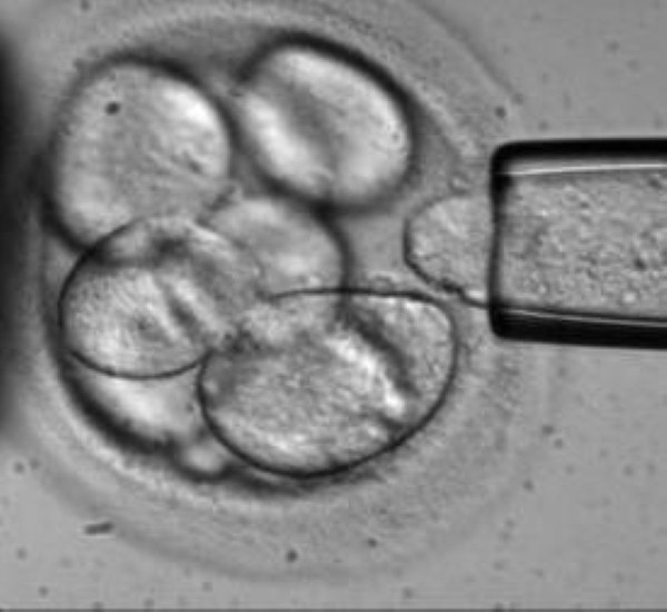
Scientists at Temple University, Philadelphia, have successfully eliminated the HIV virus from cultured human cells.

Patients affected by HIV-1 have to take medication throughout their lives to ensure they remain healthy because the virus inserts its DNA into its host's DNA. However, researchers have found a way to remove the integrated HIV-1 genes from cells permanently.

The researchers created molecular tools which they used to delete the HIV-1 proviral DNA. An enzyme known as a a nuclease and a targeting strand of RNA called a guide RNA (gRNA) were deployed to track down the viral genome and remove the HIV-1 DNA.

Once this process had taken place, the cell's natural repair mechanisms were able to take over and tie the loose ends of the genome back together.

"Since HIV-1 is never cleared by the immune system, removal of the virus is required in order to cure the disease," said Kamel Khalili, professor and chair of the Department of Neuroscience at Temple, who led the project along with Wenhui Hu, associate professor of Neuroscience.

He added that the same technique could theoretically be used against a variety of viruses.

However, the researchers must overcome a number of hurdles before the treatment is ready for patients. They need to devise a method to ensure the therapeutic agent is delivered to every affected cell and individualise treatment for for each patient’s unique viral sequences.

"We are working on a number of strategies so we can take the construct into preclinical studies," professor Khalili said. "We want to eradicate every single copy of HIV-1 from the patient. That will cure AIDS. I think this technology is the way we can do it."

According to the World Health Organisation, 34 million people around the world are currently living with HIV. The virus is particularly common in sub-Saharan African countries, such as South Africa, Zimbabwe and Mozambique.

In the UK, there were an estimated 98,400 people in the UK living with HIV at the end of 2012. With early diagnosis and effective treatment, most people infected with the virus will not go on to develop AIDS.


Categories
Microbiology

MSU team make stem cell breakthrough

Scientists at Michigan State University (MSU) have identified a gene that could make it easier to develop stem cells, which have the potential to benefit millions of people.

While the gene, which is known as ASF1A, was not discovered by the team, they determined that it is one of those responsible for cellular reprogramming – a phenomenon which is key to stem cell production as it can transform one cell into another.

"This has the potential to be a major breakthrough in the way we look at how stem cells are developed," said Elena Gonzalez-Munoz, a former MSU post-doctoral researcher and first author of the team's paper, which was recently published in the journal Science.

"Researchers are just now figuring out how adult somatic cells such as skin cells can be turned into embryonic stem cells. Hopefully this will be the way to understand more about how that mechanism works."

The scientists analysed more than 5,000 genes from a human egg, or oocyte, before discovering that ASF1A, along with another gene known as OCT4 and a helper soluble molecule, were the ones responsible for the reprogramming.

Previous research laid the groundwork for the discovery – in 2006, an MSU team identified the thousands of genes contained within the oocyte, while in 2007 Japanese scientists found that stem cells could be created without using a human egg by introducing four other genes into cells. These cells are known as pluripotent stem cells, or iPSCs.

As they are derived directly from adult tissue, iPSCs can be a perfect genetic match for a patient.

ASF1A and OCT4 genes work in tandem with a ligand, a hormone-like substance that also is produced in the oocyte called GDF9, to facilitate the reprogramming process.

Jose Cibelli, an MSU professor of animal science and a member of the team, said many more genes have yet to be discovered that play a part in the cellular reprogramming process.

The team hopes to be able to conduct further research on oocytes that will enable them to develop new, safer stem cell strategies.

Categories
Microbiology

Scientists discover ribosome ‘missing link’

Researchers at the University of California (UC) San Diego have discovered the 'missing link' in the system that enables animal cells to produce ribosomes.

The discovery could give biologists a better understanding of how to limit uncontrolled cell growth, such as cancer, that might be regulated by controlling the output of ribosomes. It will also lead to the revision of basic textbooks on molecular biology.

Ribosomes contained within each cell manufacture all of the proteins needed to build tissue and sustain life. They are responsible for a wide range of substances, including enzymes; structural molecules, such as hair, skin and bones; hormones like insulin; and components of our immune system such as antibodies.

While much time has been devoted to studying ribosomes, researchers have hitherto had little understanding of the processes underlying the formation of the proteins that are used to construct ribosomes.

Ribosomes are composed of around 80 different proteins in multicellular animals, as well as four different kinds of RNA molecules. In 1969, scientists found that two enzymes, RNA polymerase I and RNA polymerase III, are responsible for the synthesis of ribosomal RNAs.

However, they did not know whether a complementary system was also responsible for the production of the 80 proteins that make up the ribosome.

The UC San Diego researchers set out to solve this problem and discovered the missing link – the specialised system that allows ribosomal proteins themselves to be synthesised by the cell.

"We found that ribosomal proteins are synthesized via a novel regulatory system with the enzyme RNA polymerase II and a factor termed TRF2," said professor of biology and leader of the study Jim Kadonaga.  

"For the production of most proteins, RNA polymerase II functions with a factor termed TBP, but for the synthesis of ribosomal proteins, it uses TRF2."

Professor Kadonga added that the discovery of specialised TRF2-based system for ribosome biogenesis provides new opportunities for study and could potentially help to develop cancer treatments.

Categories
Microbiology

New research ‘could help combat atherosclerosis’

Researchers have identified a molecule that plays a role in exacerbating atherosclerosis and could provide a target for new therapies.

Scientists at the University of Texas (UT) Southwestern Medical Center found that a molecule known as 27HC (27-hydroxycholesterol) promotes the formation of atherosclerotic plaques, which can lead to cardiovascular disease.

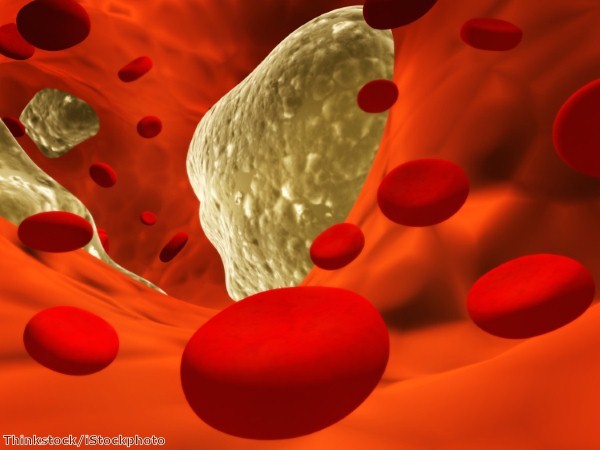
Atherosclerosis involves the build-up of lesions (or plaques) formed from lipids, such as cholesterol and fatty acids. If these rupture, they can partially or completely block blood flow, causing a heart attack or stroke.

27HC belongs to a family of molecules known as oxysterols. It is produced during the normal breakdown of cholesterol and is known to accumulate in atherosclerotic plaques.

The UT researchers discovered that 27HC promotes the formation of atherosclerotic plaques, causing a doubling in the accumulation of lipids in the arterial wall.

It achieves this through mechanisms mediated by estrogen receptors. Normally, these receptors enable estrogen to protect against the development and progression of atherosclerosis – but 27HC blocks them and prevents these beneficial effects from being realised.

"When 27HC is present, estrogen's protective effects are only observed at very high levels of the hormone," said senior author Dr Philip Shaul, holder of the Associates First Capital Corporation Distinguished Chair in Pediatrics. 

"This result may explain why hormone therapy with estrogen does not provide cardiovascular benefit in women with pre-existing atherosclerosis, in which 27HC is abundant in the vascular wall."

The researchers found 27HC triggers inflammation in the arterial wall, a key step in the establishment of atherosclerotic plaques. 

This was characterised by the exaggerated production of molecules known as cytokines that drive inflammation, as well as the enhanced attachment on the arterial wall of immune cells known as macrophages.

Macrophages then accumulate lipids (such as cholesterol) and trigger the formation of atherosclerotic plaques.

Dr Shaul said complementary therapies are needed to combat atherosclerosis, even though statins have already had dramatic impact, and targeting 27HC could help to fulfil this role.

Categories
Microbiology

New T cell therapy protects immunodeficient patients

Scientists at Technische Universitat Munchen (TUM), together with colleagues at Frankfurt, Wurzburg and Gottingen, have discovered a new method to protect patients against viruses following bone marrow transplants. 

Immune system cells are created from stem cells in the bone marrow. In diseases affecting the bone marrow, such as leukemia, degenerate cells must be destroyed using radiation or chemotherapy.

Stem cells from a healthy donor are required to replace the cells of the hematopoietic system following treatment. However, a temporary weakening of the immune system can leave patients vulnerable to infections that would not normally pose a threat.

One such agent is the cytomegalovirus (CMV), which does not cause problems in healthy human beings as specific immune cells keep the infection at bay. However, in patients with weakened defences, it can cause serious damage to the lungs or liver.

The research involved the transfer of T cells that can recognise and kill specific pathogens. The team isolated T cells from the blood of healthy donor mice and directed them against molecular elements of a bacterial species which normally causes severe infections in animals.

These cells were given to other mice, which had undergone a genetic modification so they could no longer produce immune cells of their own.  

The recipient mice were treated with bacteria after the transfer and were found to have effective immune protection, preventing them from becoming ill. 

"The most astonishing result was that the offspring cells of just one transferred donor cell were enough to completely protect the animals," first author Dr Christian Stemberger explained.

A trial was then carried out on human patients who had to undergo stem cell transplants due to a congenital immunodeficiency and leukemia, and who had contracted CMV.

T cells specifically programmed to target the CMV virus were isolated and transferred from a donor. After only a few weeks, the virus-specific cells proliferated and the number of viruses in the blood dropped.

A clinical study will now be carried out to examine the potential of the identified T cells. The scientists aim to develop innovative cell therapies using recent results and cell products created at a special TUM facility.

Categories
Microbiology

Type 1 diabetes reversal ‘could one day benefit humans’

Researchers have succeeded in reversing type 1 diabetes in mice and their efforts could help to combat the disease in humans.

Type 1 diabetes currently affects five per cent of all people with diabetes, according to the American Diabetes Association. It is usually diagnosed in children and young adults.

The incidence of the disease has increased since the mid-20th century and this could be due to under-stimulation of innate immune systems which trigger autoimmunity in children and young adults.

Individuals with type 1 diabetes do not produce enough insulin, which is central to glucose metabolism. While there is no cure for the disease, it can be controlled with insulin therapy.

Previous studies have shown that non-obese diabetic mice have defects in innate immune cells and that TLR4, a toll-like receptor, plays a protective role in preventing type 1 diabetes.

Researchers at the University of Cincinnati reversed new onset diabetes in a high percentage of newly diabetic non-obese mice using an agonistic monoclonal antibody, UT18, to boost the activity of TLR4.

"The cause of this reversal is a preservation of the endocrine pancreatic beta cells that produce insulin," explained professor William Ridgway. "These cells are preserved from the autoimmune attack which is the hallmark of type 1 diabetes."

Timing is key to reversing type 1 diabetes in mice – the disease needs to be caught at its onset, which is typically within a very short time window. This is longer in humans but is still a relatively short period from new onset to end-stage type 1 diabetes.

The new approach differs from conventional methods as the therapies in mice do not directly interact with T-cells.

Rather than targeting the adaptive immune system, Professor Ridgway's method targets the innate immune system, focusing on a receptor that is found mostly on the innate immune cells, such as dendritic cells.

"This same molecular TLR4 pathway operates in humans in many similar ways; though there are some differences, it is possible this new pathway of targeting the immune system could be tested in humans," the professor said.

Categories
Microbiology

Cancer drug raises levels of vascular-protective gene

An existing drug that is used to treat cancer patients has been found to be effective in protecting people from vascular clots. 

Bortezomib (Velcade), which is used to treat multiple myeloma, was approved for use by the US Food and Drug Association in 2012. As well as attacking cancer cells, it has been found to help prevent clot development common to many forms of the disease.

The anti-thrombotic effects of bortezomib are determined by KLF2, part of a family of Kruppel-like factors – master regulators of vascular health. These factors prevent clot formation in the body's major blood vessels.

Previous work by the researchers at Case Western Reserve University revealed that Kruppel-like factors function as nodal regulators of vascular health. This led them to surmise that bortezomib protects against thrombosis by increasing KLF levels.

Lalitha Nayak, an assistant professor of medicine, decided to test the hypothesis in the laboratory. She showed that bortezomib treatment rendered normal mice resistant to clot formation.

Next, she demonstrated that the drug specifically and potently induced KLF2 levels. Finally, she confirmed KLF2's importance by administering bortezomib to mice missing the KLF2 gene. In these animals, the drug did not have the effect of protecting them from thrombosis.

"This taught us how important KLF2 is for the ability of bortezomib to protect against thrombosis," professor Nayak said.

The results of the study could alter the management of thrombosis in patients who have a predisposition to clot formation, particularly where present treatments are ineffective.

Antiphospholipid antibody syndrome (APLS) is one such condition. Patients with the disorder have an increased risk for blood clots in both arteries and veins. It often affects young women, and there is no effective antithrombotic strategy for this group of patients.

Bortezomib protects against thrombosis and does not increase bleeding, making this drug a potential treatment alternative for APLS patients.

"Vascular clots are the number one cause of death and disability worldwide," professor Nayak said. "Our studies show that increasing KLF2 levels provides a favorable thromboprotective effect."

Categories
Microbiology

Scientists explain link between stress and heart disease

Researchers have uncovered a possible explanation for the observation that stress, emotional shock, or overexertion may trigger heart attacks in vulnerable people.

Hormones released during such events may cause bacterial biofilms on arterial walls to disperse and this could allow plaque deposits to rupture into the bloodstream.

"Our hypothesis fitted with the observation that heart attack and stroke often occur following an event where elevated levels of catecholamine hormones are released into the blood and tissues, such as occurs during sudden emotional shock or stress, sudden exertion or over-exertion" said Professor David Davies of Binghamton University, New York, an author on the study.

The research team isolated and cultured different species of bacteria from diseased carotid arteries that had been removed from patients with atherosclerosis.

They found multiple bacterial species living in the walls of every atherosclerotic (plaque-covered) carotid artery tested.

Biofilms contain communities of microbes that are resistant to antibiotic treatment and clearance by the immune system. 

When they receive certain molecular signals they disperse and release enzymes that break down the scaffolding that maintain the bacteria within the biofilm. These enzymes could dissolve the nearby tissues that prevent the arterial plaque deposit from rupturing into the bloodstream.

An experiment was conducted to test the theory by adding norepinephrine, at a level that would be found in the body following stress or exertion, to biofilms formed on the inner walls of silicone tubing. 

Professor Davies said at least one species of bacteria was able to undergo a biofilm dispersion response when exposed to norepinephrine.

The dispersal of a biofilm could trigger the sudden release of the surrounding arterial plaque and lead to a heart attack.

Managing bacteria within an arterial plaque lesion may therefore be as important as managing cholesterol, the research suggests.

Almost 160,000 people in the UK died of cardiovascular disease in 2011 and 74,000 of these deaths were caused by coronary heart disease, making it the country's biggest killer.

Categories
Microbiology

Longer telomeres linked to increased risk of brain cancers

New research led by scientists at UC San Francisco has revealed a link between common gene variants that lead to longer telomeres and an increased risk of developing deadly brain cancers known as gliomas.

Variants in two telomere-related genes known as TERT and TERC are respectively carried by 51 per cent and 72 per cent of the general population.

Telomeres are the caps on chromosome ends thought by many scientists to confer health by protecting cells from ageing.

It is thought that the benefits conferred by these variants in terms of improved cellular robustness outweigh the increased risk of developing gliomas, which are invariably fatal but relatively rare.

A massive genomic analysis of 40,000 individuals conducted at the University of Leicester found that shorter telomeres were associated with a significantly increased risk of cardiovascular disease.

The first phase of the new study involved analysing genome-wide data from 1,644 glioma patients and 7,736 healthy control individuals, including some who took part in The Cancer Genome Atlas project sponsored by the National Cancer Institute and National Human Genome Research Institute.

This confirmed the link between TERT and gliomas established in previous research and identified TERC as a glioma risk factor for the first time.

As both genes were used to regulate the action of telomerase, the enzyme that maintains telomere length, the research team analysed the Leicester data and found the two variants associated with glioma risk were also linked with greater telomere length.

Much previous research has linked longer telomeres to better health. However, as cancer cells promote their longevity by maintaining telomere length, many drug companies have searched for drugs to specifically target and block telomerase in tumors in the hopes that cancer cells will accumulate genetic damage and die.

TERT variants are also thought to play a role in lung, prostate, testicular and breast cancers, and TERC variants in leukemia, colon cancer and multiple myeloma. The research thus has a potentially much broader relevance than the glioma-related study.

Categories
Microbiology

Research identifies gene involved in Parkinson’s disease

A team of researchers at the University of California Los Angeles (UCLA) has identified a new gene involved in Parkinson's disease, potentially providing a target for drugs that could treat or cure the disorder.

Parkinson's disease involves the gradual breakdown and death of multiple neurons in the brain, leading to movement impairments, such as tremor, rigidity, slowness in movement and difficulty walking. Depression, anxiety, sleeping difficulties and dementia can also occur as a result of the illness.

Dr Ming Guo, the study team leader, associate professor of neurology and pharmacology and a practicing neurologist at UCLA, has worked on a number of studies investigating the genetic origins of the disease.

Dr Guo's team was one of two groups which found that two genes involved in inherited cases of Parkinson's disease – PTEN-induced putative kinase 1 (PINK1) and PARKIN – are also responsible for maintaining the health of mitochondria, which provide energy for the cell and are important in maintaining brain health. Mutations in these genes lead to early onset Parkinson's disease.

When these genes are working properly, they help maintain the shape of healthy mitochondria and eliminate damaged ones. Parkinson's disease can result from the accumulation of unhealthy or damaged mitochondria in neurons and muscles.

The new study found that a gene known as MUL1 (also known as MULAN and MAPL) plays an important role in mediating the pathology of the PINK1 and PARKIN.

Experiments on fruit flies and mice found that providing an extra amount of MUL1 ameliorates the mitochondrial damage due to mutated PINK/PARKIN, while inhibiting MUL1 makes the damage worse.

The team also found that removing MUL1 from mouse neurons of the PARKIN disease model leads to unhealthy mitochondria and degeneration of the neurons.

"We are very excited about this finding," said Dr Guo. "There are several implications to this work, including that MUL1 appears to be a very promising drug target and that it may constitute a new pathway regulating the quality of mitochondria."